
Abstract
Extranodal natural killer/T-cell lymphoma (ENKTL) is an Epstein-Barr virus-associated, rapidly spreading lymphoproliferative disorder with a poor prognosis. Although it usually manifests with nasal involvement, different clinical and histopathological features can be detected. In nasal form, it may be confused with mucormycosis. Identifying the appropriate biopsy site is crucial for an accurate ENKTL-nasal type diagnosis, and positron emission tomography/computed tomography (PET/CT) can facilitate this. In this paper, we share the case of a 73-year-old male who presented with nasal and skin involvement, and was diagnosed with an unfavourable prognosis using PET/CT-guided biopsy. Unfortunately, the initial biopsies were inconclusive and led to an unnecessary antimicrobial treatment. Combining fluorodeoxyglucose PET/CT images allowed us to pinpoint the biopsy location and assess the depth of tissue infiltration, resulting in a successful biopsy even in this rare case.
Keywords: Extranodal NK/T-cell lymphoma, positron emission tomography/computed tomography, skin
INTRODUCTION
Extranodal natural killer/T-cell lymphoma, nasal type (ENKTL-NT), is a rare lymphoproliferative disorder associated with Epstein-Barr virus infection. It typically occurs in the nasal cavity but can affect other areas as well. Necrosis is common (). The 5-year survival rate is 41% for the NT, but 22% for other sites. It is more common among Asians, Mexicans, and South Americans of Native American descent ().
ENKTL-NT diagnosis involves histopathological findings, immunohistochemistry, flow cytometry, and T-cell receptor (TCR) rearrangement studies (). Although biopsy improves diagnostic accuracy, selecting the most appropriate site for biopsy is challenging.
Positron emission tomography/computed tomography (PET/CT) could be helpful in diagnosis and staging, and could guide biopsy (, ). Fluorodeoksiglukoz (FDG) PET/CT plays a crucial role in the primary staging, treatment response, and follow-up of lymphomas (, ).
This article aims to share the experience of a patient with ENKTL-NT who had an atypical clinical course, a poor prognosis, and experienced a prolonged diagnostic process. Furthermore, it highlights the important role of PET/CT in diagnosis and biopsy guidance.
CASE REPORT
A 73-year-old male patient presented with persistent necrotising soft tissue infection on his right hand, forearm, and left under-eye ( and ). The patient had been presenting with lesions over the past 2-3 weeks, even after receiving empirical antibiotic treatment. The patient had no known chronic disease. The patient had experienced a wrist drop approximately 5-6 months ago; the cause could not be determined. He had been experiencing nasal congestion for a while. The patient was admitted to the department of infectious diseases and clinical microbiology; laboratory tests yielded the following results: haemoglobin: 11.1 g/dL, total leukocyte count: 4400/mm³, platelets: 114000/mm³, C-reactive protein: 5 mg/dL, erythrocyte sedimentation rate: 21 mm/h, and procalcitonin: 0.1 ng/mL. The results of the remaining biochemical and urinary tests demonstrated values within the reference range. Through the use of culture and serology, a number of infectious diseases-specifically nocardiosis, cutaneous leishmaniasis, tuberculosis, and anthrax-were successfully eliminated. Paranasal CT showed opacity in the left maxillary sinus and nasal cavity. The lesion below the eye exhibited progression over the following days, and a white lesion emerged on the hard palate (). Considering mucormycosis, liposomal amphotericin B was added to the treatment. A biopsy was taken from the patient’s palate and under the eye. The biopsy results were negative for malignancy and microorganisms. However, after 40 days of hospitalisation, a decline in the patient’s general condition was observed. An FDG PET/CT scan was conducted after 40 days to determine the potential presence of malignancy.
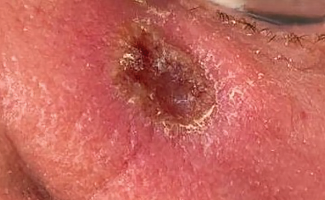
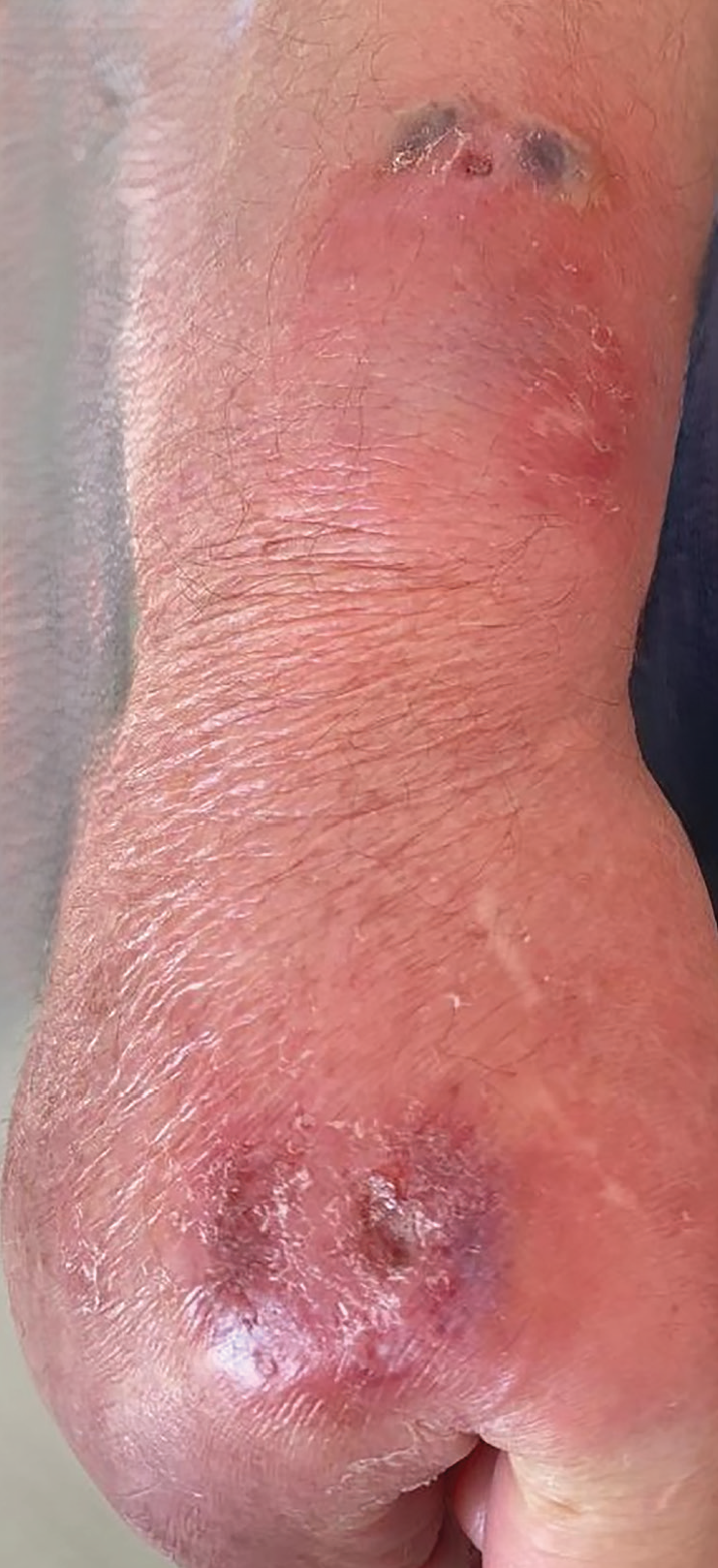
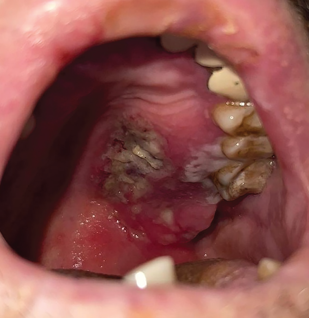
The PET/CT exam found thickening in the left maxillary region, diffuse hypermetabolic submucosal infiltration on the soft hemi-palate and parapharynx, obstructive maxillary sinusitis, and diffuse swelling on the unilateral orbital floor [maximum standard uptake value (SUVmax): 35.4]. Similar involvement with hypermetabolic subdermal infiltration was seen in the right forearm and hand (SUVmax: 15.6). A few bilateral cervical lymph nodes (SUVmax: 6.5) were noted, as well as an axillary and a left hilar lymph node (SUVmax: 4.2) were noted. In addition, the spleen was enlarged, and the liver/spleen metabolic ratio was increased ().
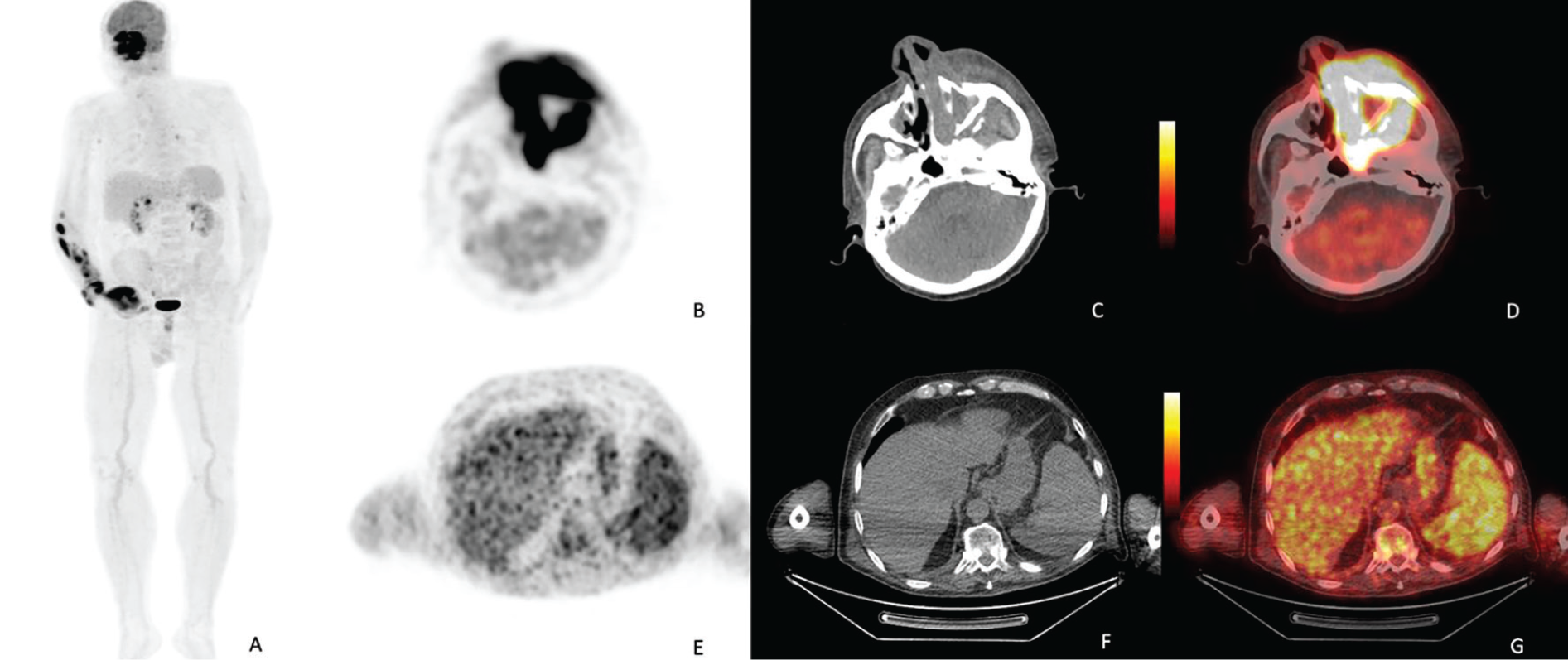
PET: Positron emission tomography, CT: Computed tomography
No concerning surface lymph nodes were found; therefore, a PET/CT-guided biopsy was conducted. The biopsy depth was determined by analysing forearm photos, emission images, and CT sections. The extensor sites of the hand and wrist were chosen for dermal and subdermal sampling.
Lymphoid cell infiltration with angiodestruction was detected in the adipose tissue of the hand and wrist. Immunostaining showed that the neoplastic cells were positive for CD3, CD5, CD7, CD56, T-cell intracellular antigen 1, granzyme B, and Epstein-Barr encoding region (EBER). However, they were negative for CD20, CD4, CD8, CD30, PD-1, TCR beta, and TCR gamma/delta, indicating the presence of ENKTL ( and ).
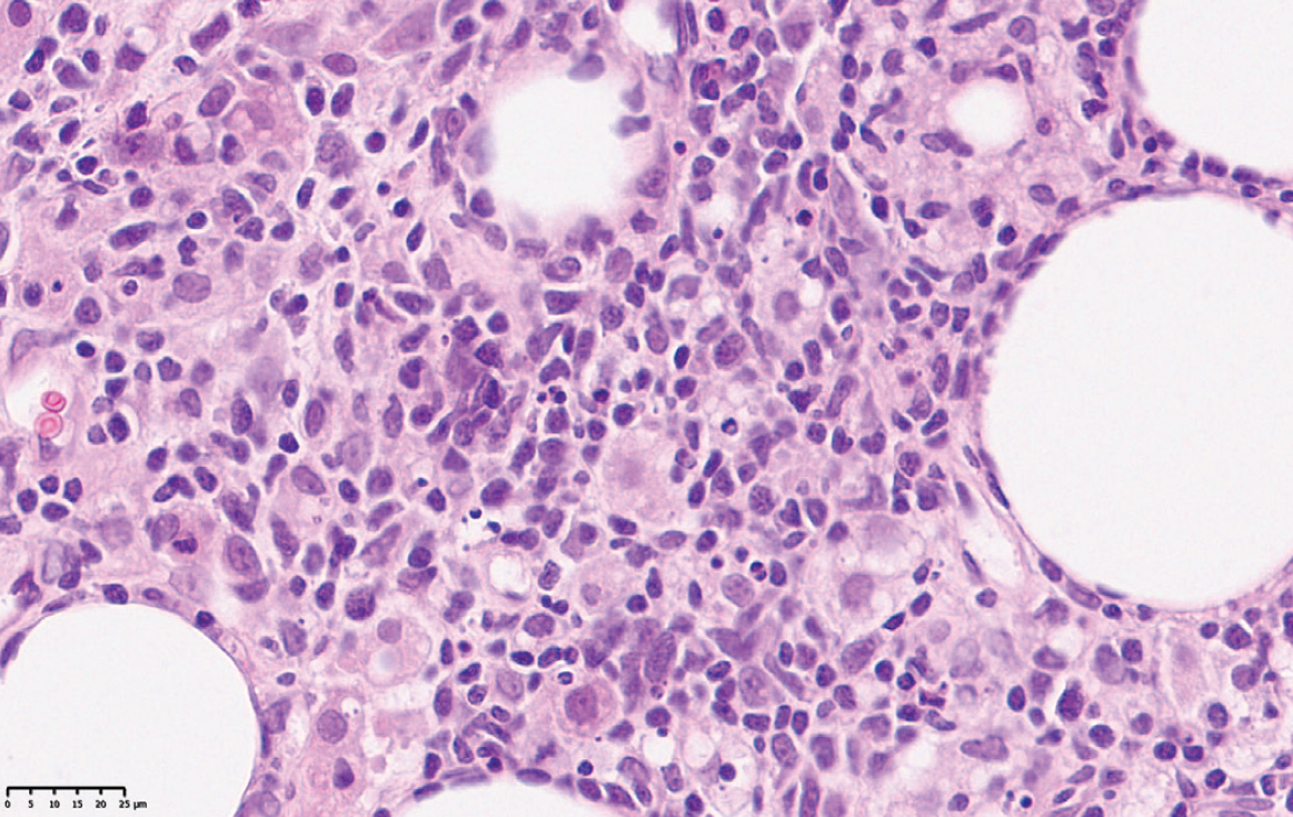
H&E: Hematoxylin and eosin
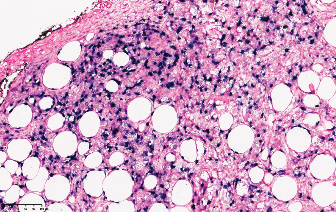
The patient was referred to the haematology clinic for chemotherapy, but unfortunately, he passed away due to cardiac arrest.
DISCUSSION
Patients diagnosed with ENKTL-NT typically exhibit a low incidence of skin lesions occurring beyond the facial region. Skin lesions, whether singular or multiple, can often be mistaken for infections such as cellulitis, abscesses, and infective panniculitis. The histopathological patterns of cutaneous involvement in ENKTL-NT share similarities with other lymphoma variants, dermatitis, and infectious processes. Notably, angiocentricity, angioinvasion, and epidermotropism are common histopathological findings (). Accurate diagnosis is often challenging due to extensive angioinvasion and necrosis, requiring multiple biopsies (). In our case, skin lesions provided misleading clinical information, and the initial biopsy samples were non-diagnostic.
ENKTL-NT is characterised by bloody rhinorrhoea and nasal obstruction, as well as systemic symptoms such as prolonged fever and weight loss (). Although the patient in our case presented with nasal obstruction, B symptoms were absent, except during the last period. The literature suggests that having two or more extranodal sites and being over 60 years old are factors that can lower overall survival rates (). Unfortunately, in our case, the patient was diagnosed with ENKTL-NT at an advanced age, and multiple involvements in the skin, soft tissue, spleen, and liver indicated a poor prognosis. EBER positivity is also an important diagnostic indicator (). In our case, the patient exhibited EBER positivity accompanying tissue lesions on pathological examination.
Several prognostic models have been studied for ENKTL-NT. PET/CT parameters are used in one of these models (). Li et al. () reported that the patients in the high SUVmax group had significantly inferior progression-free survival and overall survival rates when the optimal SUVmax cut-off value was determined to be 9.5. SUVmax levels were very high in our patient, especially in the face and arm involvement. Moreover, PET/CT scans can play a significant role in guiding biopsies. We used PET/CT to determine the optimal site for biopsy in our patient.
In conclusion, ENKTL-NT remains a concerning lymphoma in terms of the diagnosis, follow-up, and treatment of patients. It is important to distinguish ENKTL-NT from other medical conditions such as necrotising soft tissue infections, mucormycosis, and dermatitis. In ENKTL-NT patients, unnecessary diagnostic and treatment procedures lead to delays. PET/CT may help guide invasive procedures accurately and prevent unnecessary biopsies, resulting in patients being evaluated less invasively and more effectively.
Footnotes
Ethics
Informed Consent: The written informed consent form was obtained.
Authorship Contributions
Surgical and Medical Practices: Z.K., M.B., A.A., M.K., N.A., Ş.A.B., G.Y.T., C.B. Concept: Z.K., M.B., A.A., M.K., N.A., Ş.A.B., G.Y.T., C.B., Design: Z.K., M.B., A.A, M.K., N.A., Ş.A.B., G.Y.T., C.B., Data Collection or Processing: Z.K., M.B., A.A., M.K., N.A., Ş.A.B., G.Y.T., C.B., Analysis or Interpretation: Z.K., M.B., A.A., M.K., N.A., Ş.A.B., G.Y.T., C.B., Literature Search: Z.K., A.A.., M.K. Writing: T Z.K., M.B., A.A., M.K., N.A., Ş.A.B., G.Y.T., C.B.
Conflict of Interest: The authors declared no conflict of interest
Financial Disclosure: The authors have not declared any financial support.
References
Murga-Zamalloa C, Inamdar K. Classification and challenges in the histopathological diagnosis of peripheral T-cell lymphomas, emphasis on the WHO-HAEM5 updates. Front Oncol. 2022; 12: 1099265.
Kim TM, Lee SY, Jeon YK, Ryoo BY, Cho GJ, Hong YS, et al. Clinical heterogeneity of extranodal NK/T-cell lymphoma, nasal type: a national survey of the Korean Cancer Study Group. Ann Oncol. 2008; 19: 1477-84.
He X, Gao Y, Li Z, Huang H. Review on natural killer/T-cell lymphoma. Hematol Oncol. 2023; 41: 221-9.
Jeong SH. Extranodal NK/T cell lymphoma. Blood Res. 2020; 55: S63-71.
Geng H, Li J, Zhang W. Comparison of 18F-FDG PET/CT and conventional methods in diagnosing extranodal natural killer/T-cell lymphoma. Heliyon. 2023; 10: e23922.
Li H, Shao G, Zhang Y, Chen X, Du C, Wang K, et al. Nomograms based on SUVmax of 18F-FDG PET/CT and clinical parameters for predicting progression-free and overall survival in patients with newly diagnosed extranodal natural killer/T-cell lymphoma. Cancer Imaging. 2021; 21: 9.
Ngamdamrongkiat P, Sukpanichnant S, Chairatchaneeboon M, Khuhapinant A, Sitthinamsuwan P. Cutaneous ınvolvement of extranodal NK/T cell lymphoma, nasal type, a clinical and histopathological mimicker of various skin diseases. Dermatopathology (Basel). 2022; 9: 307-20.
Liang JH, Ding CY, Gale RP, Wang L, Xu J, Qu XY, et al. Prognostic value of whole-body SUVmax of nodal and extra-nodal lesions detected by 18F-FDG PET/CT in extra-nodal NK/T-cell lymphoma. Oncotarget. 2017; 8: 1737-43.